Anatomy of Your Child’s Respiratory System
The respiratory system carries air in and out of the lungs. When your child breathes in, oxygen-rich air flows into the lungs. This oxygen is sent to all the body’s cells to use for energy and growth. When your child breathes out, waste gases (carbon dioxide) flow out.
How breathing works
When your child breathes in, air fills the airways in the lungs. Oxygen-rich air reaches the balloon-like air sacs at the end of the airways. These sacs are called alveoli. Oxygen passes into the blood vessels around the sacs. The blood then carries the oxygen to all parts of the body. As the body uses oxygen, it makes a waste gas (carbon dioxide). The blood carries that back to the lungs. When your child breathes out, carbon dioxide leaves the body through the airways, windpipe, and mouth or nose.
Parts of the respiratory system
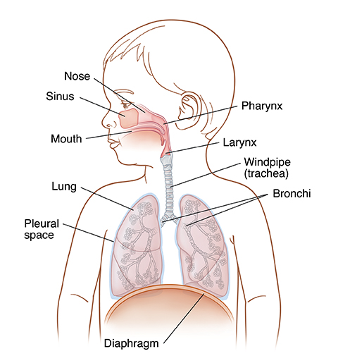
A child’s and an adult's respiratory system are a lot alike. But some structures have a different size or position. For example, a baby's tongue takes up more space in the mouth than an adult's tongue. And a baby's larynx is in a higher part of the neck than it is for an adult.
-
The mouth and nose. These are the openings through which air enters and exits the body.
-
Sinuses. These are air-filled chambers in the bones of the face. They help keep the nose moist and free of dust and bacteria.
-
The pharynx. This is the cavity behind the mouth.
-
The larynx. This is the upper part of the windpipe, which contains the vocal cords.
-
The windpipe (trachea). This is a pathway for air to enter and exit the lungs.
-
The lungs. These 2 organs are made of spongy tissue. They have 5 parts (lobes), 3 in the right lung and 2 in the left. The lungs allow the body to receive oxygen and get rid of carbon dioxide.
-
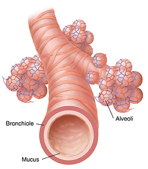
Bronchi (right and left). These connect the trachea to the two lungs. Each bronchus branches into smaller tubes called bronchioles. Bronchioles (airways) are stretchy “branches” that move air all over the lungs. Bands of muscles surround each bronchiole. Bronchioles get smaller as they go deeper into the lungs.
-
Alveoli. These are clusters of balloon-like air sacs at the ends of the airways.
-
Blood vessels. These are tubes that carry blood to the lungs and throughout the body. Tiny blood vessels surround the air sacs, allowing an exchange of oxygen and carbon dioxide.
-
The pleural space. This is an area between the lungs and chest wall, lined on both sides by tissue called pleura.
-
The diaphragm. This is a muscle in the belly that helps with breathing.
-
Mucus. This is a sticky substance made by cells in the lining of the airways. It traps dust, smoke, and other particles from air breathed in.
-
Cilia. These are tiny hairs on the cells of the airway lining that are coated with sticky mucus. They trap germs and foreign particles that enter from air breathed in. They then sweep them up to the nose or mouth. From there, mucus gets swallowed, sneezed, or coughed out.
Online Medical Reviewer:
Amy Finke RN BSN
Online Medical Reviewer:
Jessica Gotwals BSN MPH
Online Medical Reviewer:
Liora C Adler MD
Date Last Reviewed:
7/1/2022
© 2000-2024 The StayWell Company, LLC. All rights reserved. This information is not intended as a substitute for professional medical care. Always follow your healthcare professional's instructions.